What’s involved in an eye examination?
History & SymptomsSo that we can tailor the examination to your needs we start by asking some questions to find out why you have come to see us and if you are experiencing any problems with your eyes or vision. We will ask about any previous eye problems &/or treatments, make a list of any medications or supplements that you are taking and ask if any of your family relations have had any eye diseases.
The information we get from this chat directs what testing needs to be done. The rest of the examination will contain many of the following elements:
Vision Testing
First of all we want to know how well you see without any spectacles or contact lenses.
Refraction
Using the latest computer and subjective testing we determine if you need help with your vision, if this is the case we will advise the best spectacle lens correction for your eyes. Or if you currently wear glasses whether an update in prescription is required.
Binocular
A series of tests to check how well the muscles that control your eye movements work and if any adjustments need to be made to the spectacle lenses to help your binocular vision.
Eye exercises may also be part of the solution.
Health of Eyes
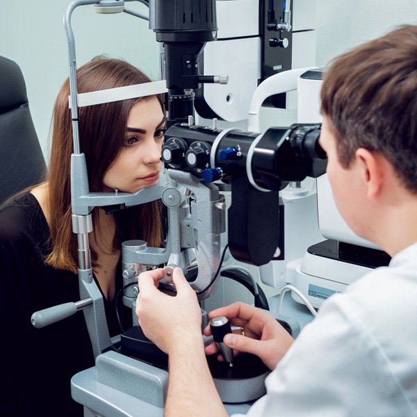
Anterior Eye ExaminationHere we check the health of the front of your eyes. This includes looking at your eye lids, the white and clear outer portions, the iris, the pupils, your crystalline or intra ocular lenses and the space between the iris and the cornea.
This part of the eye is prone to UV and traumatic damage so we use a machine called a slit lamp to help highlight any damage.
Posterior Eye Examination
Here we look through your pupil to check the health of the inside of your eyes.
The traditional way to do this is with a hand held torch called an ophthalmoscope but we take photos of the inner most surface of the retina using a digital retinal camera. This provides a wider, clearer view and we attach the images to your personal file. Comparing your most recent retinal images with older ones means that we are best placed to detect early changes in your eye health.
Depending on your symptoms and the size of your pupils we may need to put drops in your eyes to enlarge the pupils and allow a clearer, wider examination.
A 3D test called an OCT is used if the retina looks damaged or to better detect damage caused by disease or medication toxicity. It is like an MRI scan of your eyes and allows examination of many of the layers of the retina. This has revolutionised our ability to detect very early signs of eye diseases such as glaucoma, macular degeneration, diabetes and vascular disease.
Binocular Ophthalmoscopy and slit lamp provide a much wider 3D assessment of your peripheral retina and/or vitreous.
Intraocular Pressure
This measurement is taken using a machine called a tonometer of which we have various types.Glaucoma and iritis are two diseases which can alter the fluid pressure of your eye so it is good to be able to compare current and older readings. Certain medications (steroids) can also cause this.
Corneal thickness
Corneal thickness may be measured to look for disease or allow us to more accurately assess the intraocular pressure reading. This is measured using a device called a pachymeter.
Corneal Topography
Corneal topography is a non-invasive imaging technique for mapping the surface curvature of the cornea. It is used to help with some contact lens fitting and to check for and monitor progress of eye diseases such as keratoconus.
Visual Field Testing
Many people know that glaucoma reduces peripheral vision and macular degeneration affects central vision but commonly used medications, vascular and brain disease can also alter your visual fieldCentral vision is tested using an Amsler grid and we use frequency doubling technology in the form of a Humphrey Matrix Perimeter to carefully measure the sensitivity of side vision.